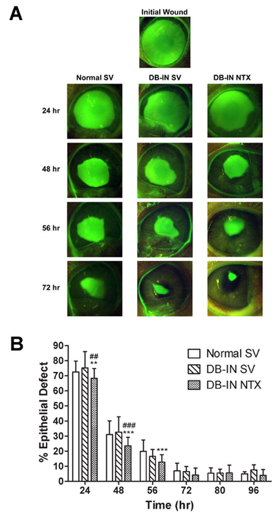
Figure 3.

Photomicrographs (A) and areal measurements (B) of Normal and insulin controlled diabetic rabbits (DB-IN) receiving either sterile vehicle (SV) or 10−4 M NTX. (A) Eyes were stained with fluorescein and photographs taken at 0 (Initial Wound), 24, 48, 56, and 72 h after abrasion. (B) Residual epithelial defect (means ± SEM; n = 10–15 animals/treatment group) as percentage of the original wound. Significantly different from Normal SV rabbits at p<0.01 (**) and p<0.001 (***), and from DB-IN SV group at p<0.01 (##) and p<0.001 (###).
