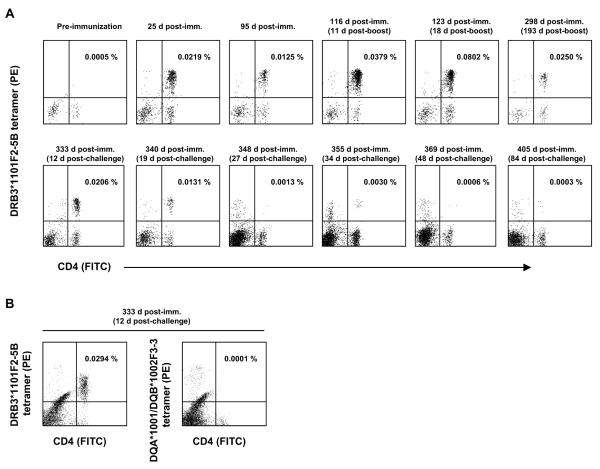
Figure 2.
Enumeration of antigen-specific CD4+ T cells in peripheral blood of animal 3959 before and after immunization and A. marginale infection. A, Cryopreserved PBMC were stained with the DRB3*1101-F2-5B tetramer at the indicated time points and analyzed by FACS. Dot plots indicate the percentage of tetramer+ CD4+ cells after anti-PE magnetic bead selection. The upper panel shows cells obtained before and after immunization and after the peptide boost on day 105. The bottom panel shows cells obtained 12-84 days following A. marginale challenge. B, Cryporeserved PBMC obtained 12 days post-challenge were thawed and stained with either the DRB3*1101F2-5B tetramer or an unrelated DQA*1101/DQB*1102F3-3 tetramer after anti-PE magnetic bead selection. The numbers in the upper right quadrants are the calculated percentage of tetramer+ CD4+ T cells of the total CD4+ T cells in the samples.

