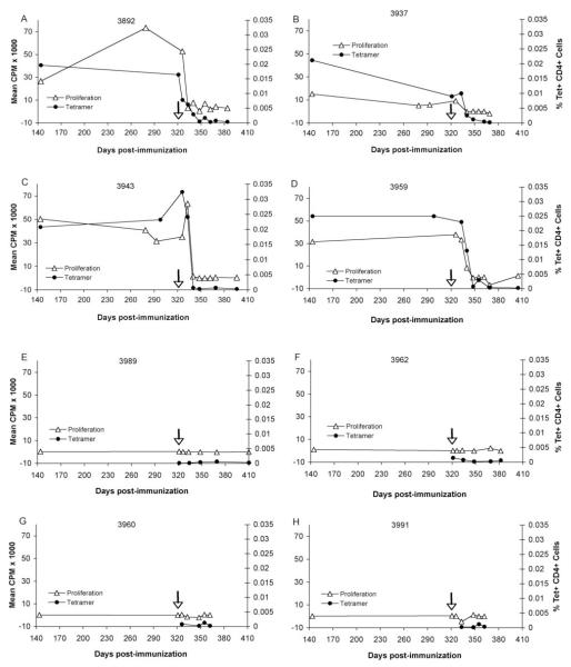
Figure 4.
T lymphocyte proliferation and enumeration of tetramer+ CD4+ cells in PBMC from vaccinates and controls before and after A. marginale infection. PBMC were obtained on day 141 post-immunization and at select time points thereafter and following challenge on day 321 (arrow). Cryopreserved PBMC were stained with the tetramer and analyzed by FACS. A-D, Results are presented for all vaccinates. E-H, Results are presented for all control animals. For proliferation assays, lymphocytes from animals 3960 and 3991 were cryopreserved, whereas lymphocytes remaining animals were fresh. The data represent the percentages of tetramer+ CD4+ cells of the total CD4+ T cells (black circles) or the mean CPM of triplicate cultures with 1 μg/ml peptide F2-5 after subtracting the background mean CPM in response to medium (white triangles).

