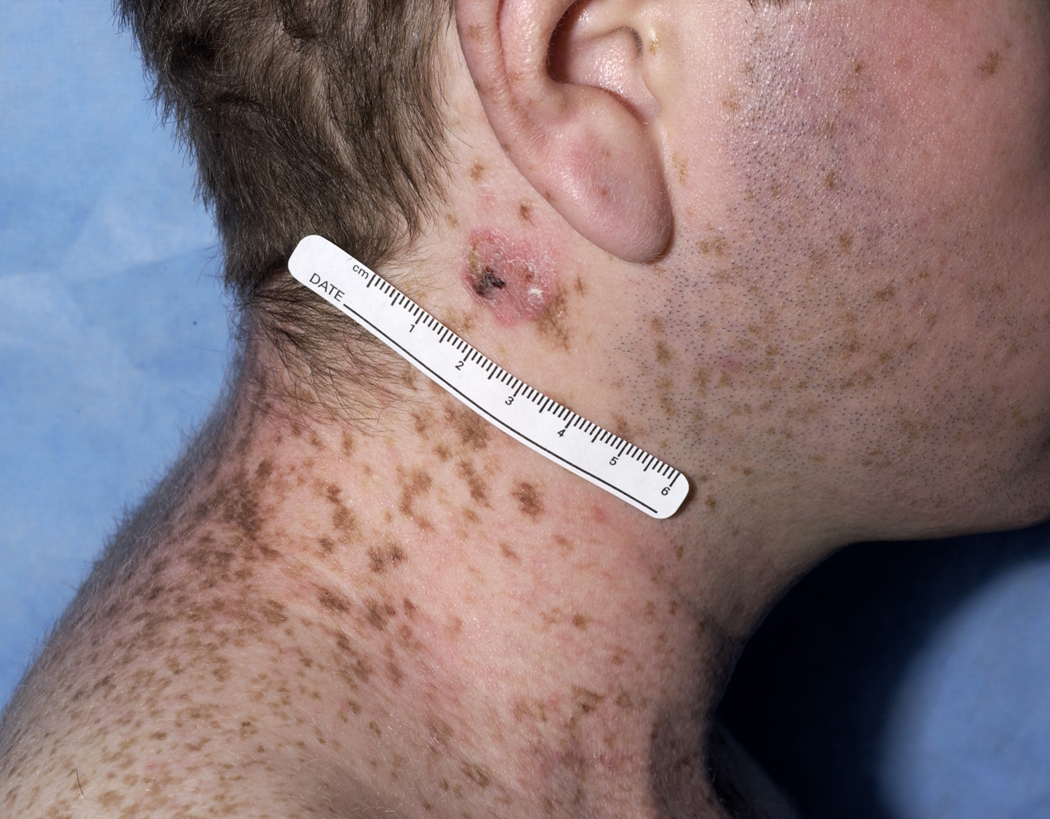
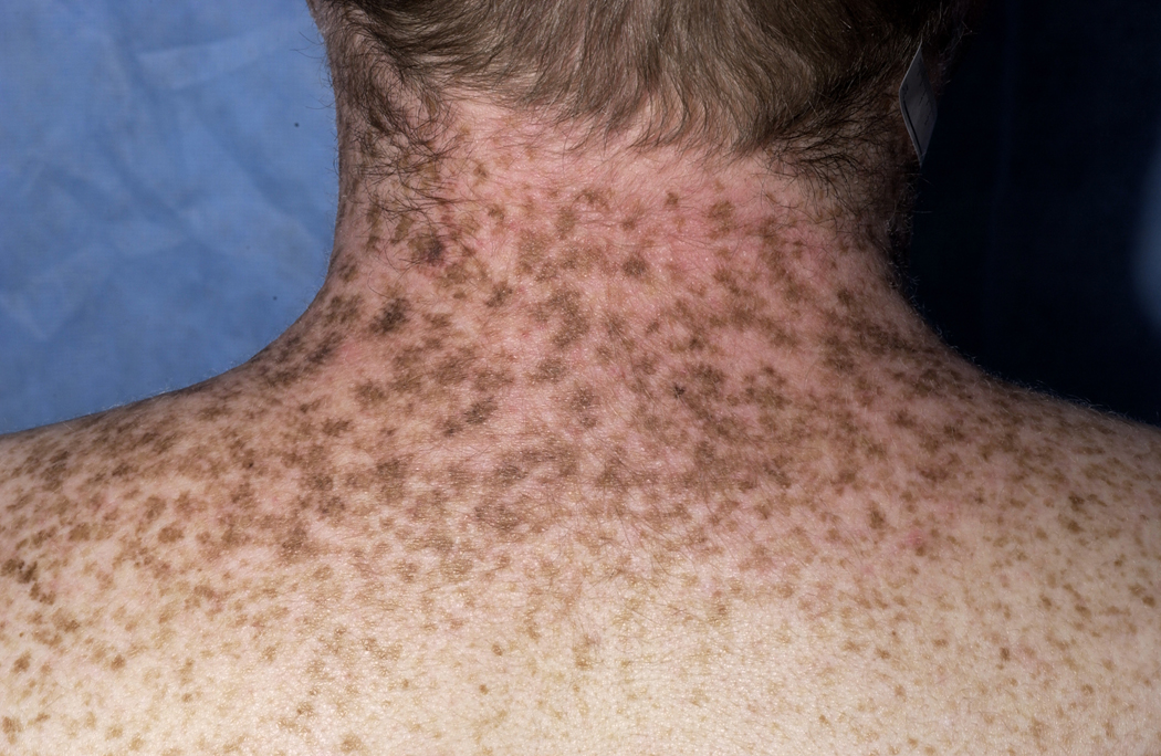
FIGURE 1. Cutaneous manifestations of long-term treatment with voriconazole.
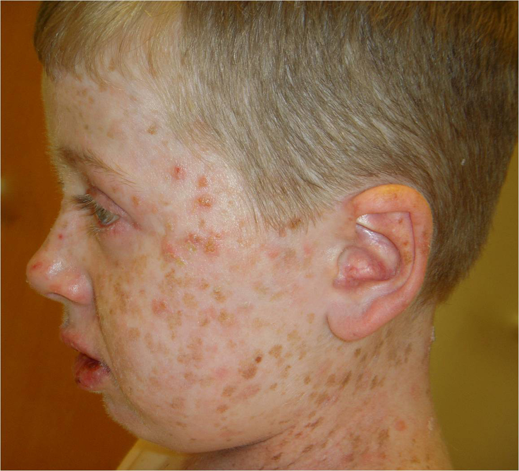
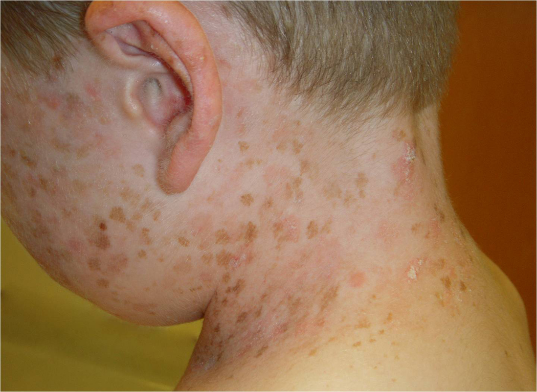
A. Nine year-old boy with extensive lentigo formation and multiple actinic keratoses on the head and neck following three years of treatment with voriconazole (Pt 1).
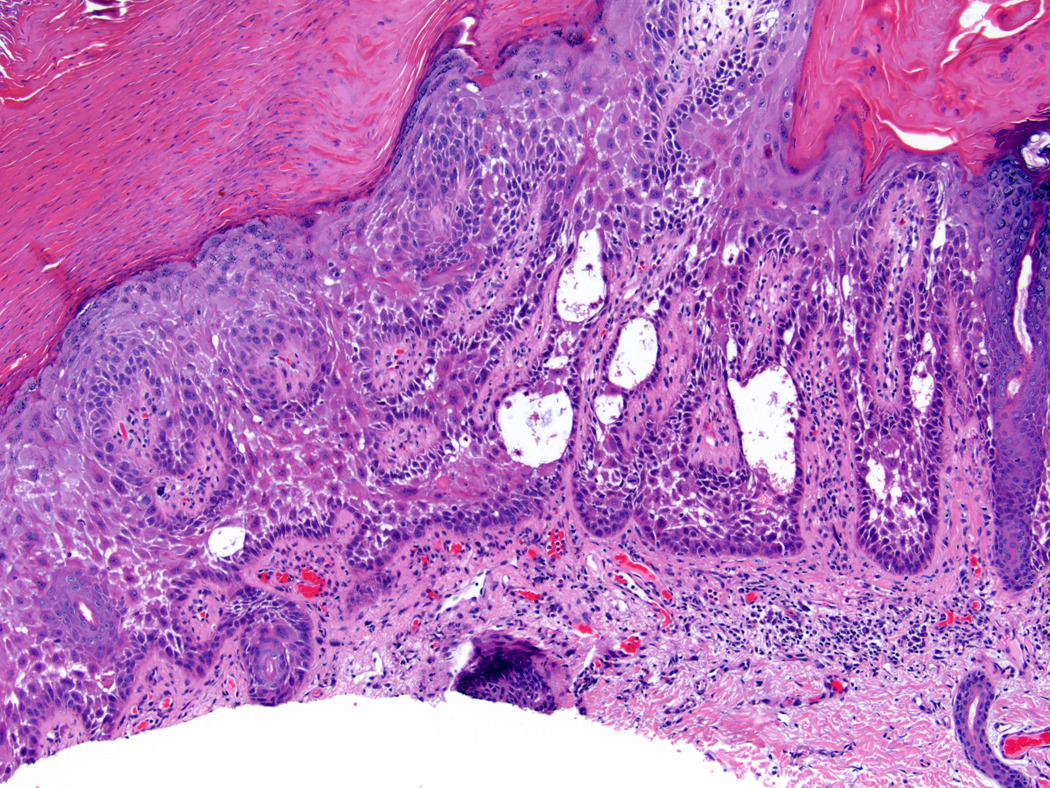
B. Hyperkeratotic lesion with erythema on the left neck of Pt 1. Histology demonstrated acanthotic solar keratosis with acantholysis, bordering on superficially invasive SCC.
C. Histology of lesion on left neck (Figure 1B) identified acanthotic solar keratosis with acantholysis. This lesion bordered on superficially invasive squamous cell carcinoma (hematoxylin and eosin, original magnification 100×).
D. 22 year-old male with long-standing HIV infection with SCC on left lateral neck following 15 months of voriconazole therapy (Pt. 3). Chronic phototoxicity with erythema and dark brown lentiginous pigmentation is present on the head and lateral neck.
E. Similar erythema and pigmentation resembling xeroderma pigmentosum is present on the photo-exposed surfaces of the posterior neck and upper extremities (Pt. 3).
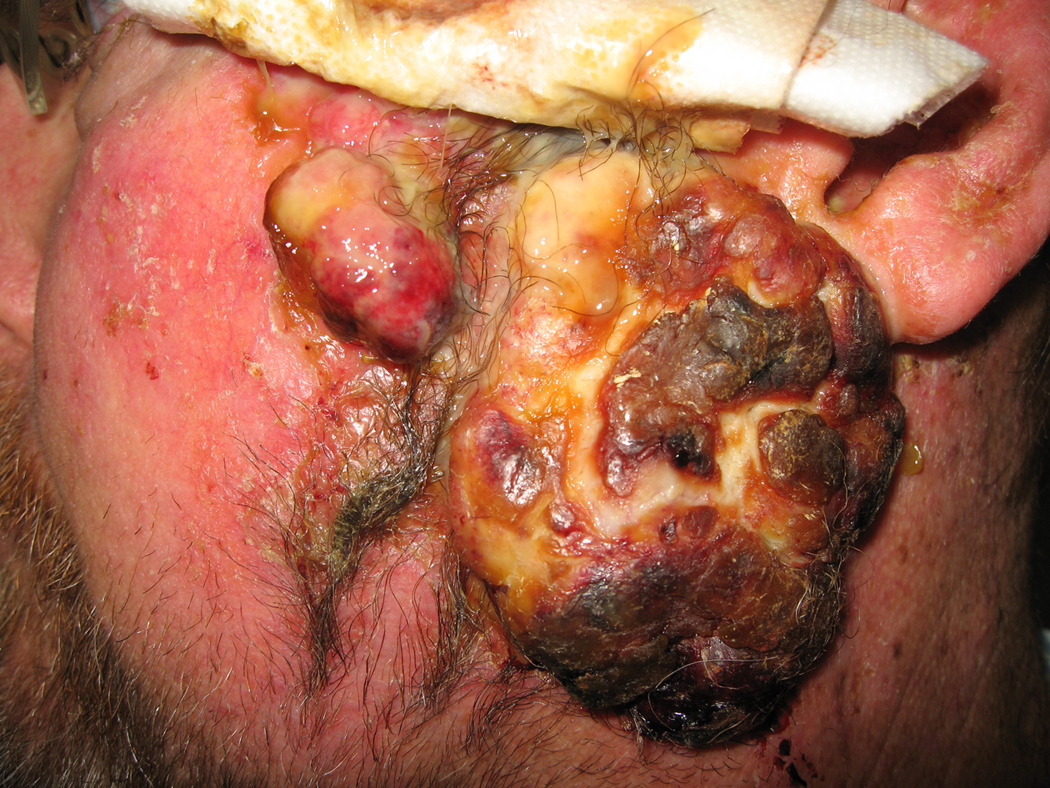
F. 46-year old male (Pt. 5) following four years of treatment with voriconazole with recurrent SCC on the left preauricular cheek. The patient subsequently died of metastatic disease.
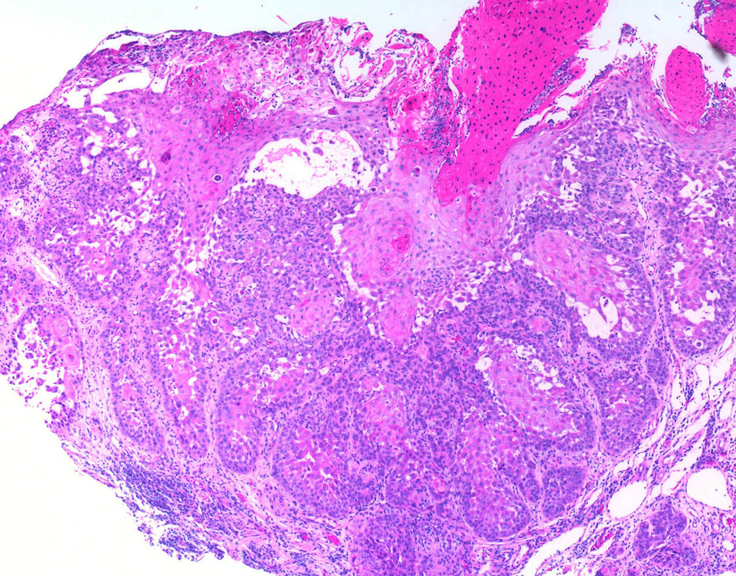
G. Histology of SCC from right cheek (Pt. 5) reveals deeply invasive SCC with keratinocyte pleomorphism, representative of the multiple SCCs that developed in this patient (hematoxylin and eosin, original magnification 40×).

