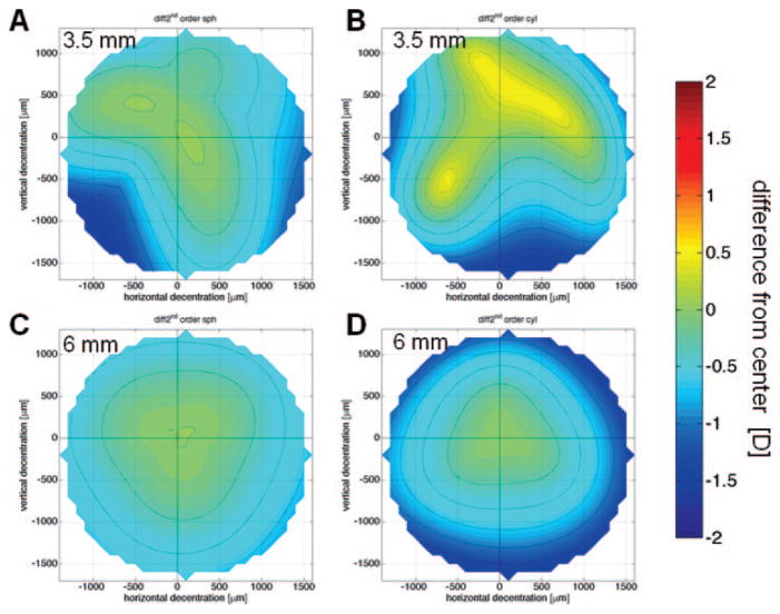
Figure 1.
Second-order refraction change decentration maps for the right eye of cat 5-005. (A) Change in sphere, 3.5-mm PD. (B) Change in cylinder magnitude, 3.5-mm PD. (C) Change in sphere, 6-mm PD. (D) Change in cylinder magnitude, 6-mm PD. The center (crosshair) is set to zero; dotted lines: 0.25-D steps.

