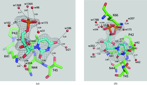
Figure 2.
View of uridine 5′-monophosphate in the pyrimidine-binding site of RNase A. Electron density from F o − F c maps calculated with the respective U5P omitted from the structure-factor calculations are shown contoured at 2.5σ. (a) U5P bound to molecule A of RNase A. (b) U5P bound to molecule B of RNase A showing the disordered phosphate group. Hydrogen-bonding interactions are shown as dashed lines with distances given in Å.

