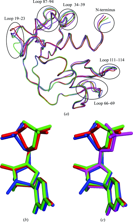
Figure 5.
(a) Superposition based on Cα atoms of the four independently determined structures of RNase A in complex with uridine 5′-monophosphate. Highly variable regions are identified. (b) U5P molecules from 3dxg and the B molecule of the present study. Relative positioning is based on the superpositioning that produced (a). (c) U5P from all four complexes. In all three images, U5P-A and U5P-B from 3dxg are shown in green and blue, respectively, and U5P-A and U5P-B from the present study are shown in magenta and red, respectively.

