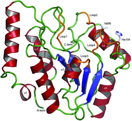
Figure 2.
Ribbon illustration of the crystal structure of vcUNG. α-Helices are illustrated in red, β-strands in blue and loop regions in orange. Loops involved in the detection and removal of uracil in DNA are marked. Loop1 is the water-activating loop (64-DPYH-67), Loop2 is the Pro-rich loop (84-KTPPS-88), Loop3 is the Gly-Ser loop (165-GS-166) and Loop4 is the Leu loop (187-HPSPLSAH-194). The sites of the mutations Val90 and His194 are also shown.

