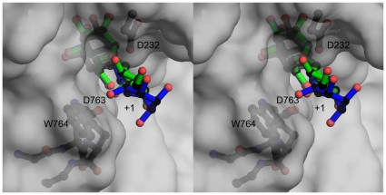
Figure 6. Substrate specificity in SpGH38.
An overlap of the Drosophila GH38 α−mannosidase II complexes [15] with α−1,3 (green bonds) and α−1,6 linked ligands (blue bonds) with the SpGH38 structure (grey surface) focussing on the +1 subsite (the -1 subsites are essentially identical, Fig. 5A). Features which may contribute to 1,3 specificity include the position of W764, the interactions afforded by D763 and the tightness of the “sphincter” formed by D763 and the catalytic acid/base D232. The figure is shown in divergent (“wall-eyed”) stereo.

