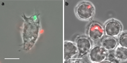
Fig. 1.
Detection of meronts and phagocytosed spores. a Differential immunofluorescence staining was applied to distinguish internalized from extracellular spores. J774 cells were cultured on cover slides, incubated with E. cuniculi spores and fixed 20 h later with 4% paraformaldehyd in PBS for 30 min. Samples were washed twice with PBS and incubated with mAb 11A1 (1:100 in PBS), which detects the spore wall protein SWP1 (Bohne et al. 2000), followed by incubation with a Cy3-conjugated anti-mouse IgG (1:500 in PBS). Afterwards, cells were permeabilized in 0.25% TritonX100/PBS for 20 min. The incubation step with mAb 11A1 was repeated, followed by IgG detection with a Cy2-conjugated anti-mouse IgG (1:300 in PBS). Fluorescence microscopy revealed a red color (Cy3) for extracellular spores and a green color (Cy2) for intracellular spores. b Detection of meronts inside parasitophorous vacuoles. Samples were fixed and permeabilized as described above and incubated with mAb 6G2, which recognizes an antigen expressed in meronts (Fasshauer et al. 2005), followed by IgG detection with a Cy3-conjugated anti-mouse IgG. Size bars are 10 µm

