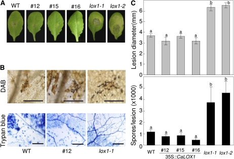
Figure 11.
Disease development on Arabidopsis wild-type (WT), CaLOX1-OX transgenic, and lox1 leaves inoculated with A. brassicicola. Leaves of 4-week-old plants were challenged with 10-μL droplets containing 5 × 10⁴ mL−1 fungal spores. A, Disease symptoms. B, Diseased leaves stained with DAB and trypan blue. Microscopic images show stained fungal structures and damaged plant cells. Bars = 0.05 mm (DAB) and 0.15 mm (trypan blue). C, Quantification of disease development. Top, average diameter of lesions caused by A. brassicicola. The lesion sizes are averages of 30 lesions per line. Lesion diameter is presented ± sd. Bottom, average numbers of newly formed spores per lesion. Spores on 10 inoculated leaves per line were counted and are presented as means ± sd. Different letters indicate significant differences as determined by the lsd test (P < 0.05) in three independent experiments. [See online article for color version of this figure.]

