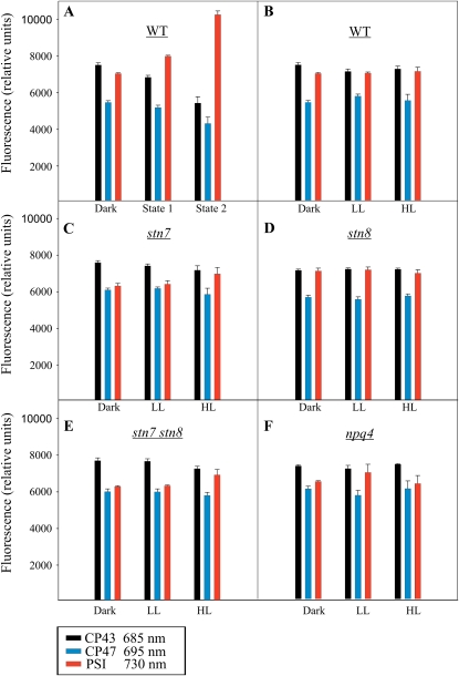
Figure 2.
The 77 K chl a fluorescence emitted from CP43 (685 nm), CP47 (695 nm), and PSI (730 nm). The 77 K chl a fluorescence was recorded in the wild type (WT; B), stn7 (C), stn8 (D), stn7 stn8 (E), and npq4 (F) after 16 h of dark incubation and subsequent 1-h exposure to 30 or 1,000 μmol photons m−2 s−1. Wild-type plants were also exposed to far-red light (state 1) and red light (state 2) for 1 h following the 16-h dark incubation (A). All thylakoid samples were prepared in the presence of 10 mm NaF and diluted to 10 μg chl mL−1 before the measurements. Data are means of three independent measurements, and error bars represent sd. [See online article for color version of this figure.]

