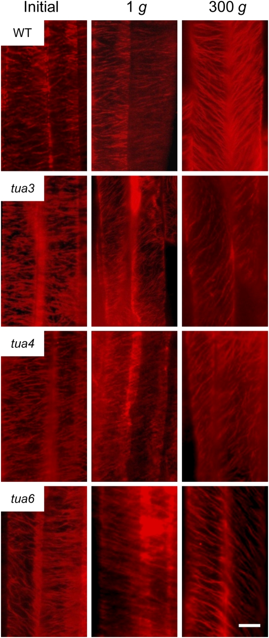
Figure 5.
Immunofluorescence images of cortical microtubules in hypocotyls of Arabidopsis tubulin mutants. Wild type (WT) and tubulin mutants were grown as in Figure 1. Epidermal cells 10 to 12 were stained as described in “Materials and Methods.” Typical examples of two adjacent cells with distinct orientation of cortical microtubules are shown. The bar denotes 10 μm.

