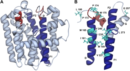
Figure 11.
Three-dimensional ribbon model of the complete structure of wild-type PaIDS1 (A) and the substrate-binding region (B) constructed with reference to the avian FPP synthase crystal structure (Tarshis et al., 1994). Depicted are the α-helices D (amino acids 154–181), F1 + F2 + G (amino acids 222–277), and H (amino acids 309–313). Helices that are part of the predicted substrate-binding pocket are colored in blue (helices D + F), and the DDxxD motifs (amino acids 170–176 and 309–313) are indicated in red. Residues used for mutagenesis that are visible in this diagram are shown in turquoise. The model was displayed with the program UCSF Chimera (National Centre for Research Resources).

