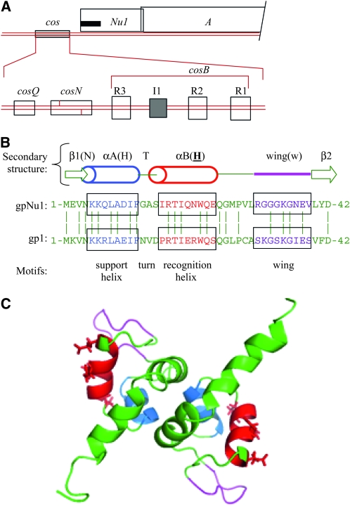
Figure 1.—
The cos and terminase region of the λ-chromosome. (A) (Top) Map of cos and the terminase-encoding Nu1 and A genes. The black bar indicates the location of the winged helix-turn-helix DNA-binding motifs in the N-terminal domain of gpNu1. (Bottom) cos subsites: cosQ is required for termination of DNA packaging; cosN is the site where the large terminase subunit, gpA, introduces staggered nicks to generate the cohesive ends of virion DNA molecules; and cosB contains the gpNu1-binding sites R1, R2, and R3 along with the IHF-binding site I1. (B) (Top) Schematic of gpNu1 residues 1–42, including the support (blue) and recognition (red) α-helixes and the wing loop (magenta). β1 and β2 are short β-strands flanking the DNA-binding elements. (Bottom) Sequences are a comparison of residues of λ's gpNu1 and phage 21's gp1, with conserved resides indicated by vertical lines. Note that the recognition helixes of gpNu1 and gp1 differ by four residues, all likely solvent-exposed (Becker and Murialdo 1990; de Beer et al. 2002). (C) Three-dimensional structure of the winged helix-turn-helix-containing, N-terminal domain of gpNu1 (residues 1–68) (de Beer et al. 2002). Side groups of solvent-exposed residues of the recognition helix are displayed. Color coded as in B.

