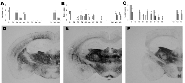
Figure 4.
Lesion profiles and regional distributions of atypical proteinase K–resistant prion protein (PrPres) in the brain of porcine PrP transgenic mice infected, either in 1st passage (white column) or in 2nd passage (black column) with cattle bovine spongiform encephalopathy (BSE) (panels A and D), sheep BSE (panels B and E), or atypical scrapie (panels C and F) agents. A–C) Lesion scoring of 9 areas of gray matter (G) and white matter (W) in mice brains: dorsal medulla (G1), cerebellar cortex (G2), superior colliculus (G3), hypothalamus (G4), medial thalamus (G5), hippocampus (G6), septum (G7), medial cerebral cortex at the level of the thalamus (G8) and at the level of the septum (G9), cerebellum (W1), mesencephalic tegmentum (W2) and pyramidal tract (W3). Error bars indicate SE. D–F) Histoblots of representative coronal sections at the level of the hippocampus.

