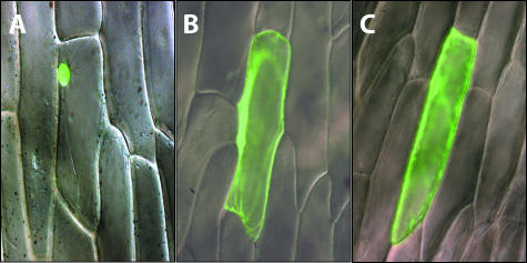
Figure 3.
Intracellular localization of GFP-AtPER1 protein fusion. Merged differential interference contrast light micrographs and fluorescence micrographs of GFP fusion constructs transiently expressed in onion epidermal cells after particle bombardment. A, 35S::HP1-GFP. The GFP fluorescence signal is only found in the nucleus. B, 35S::GFP. GFP fluorescence signal is found in the cytoplasm and the nucleus. C, 35S::GFP-ATPER1. GFP fluorescence signal is found in the cytoplasm and in the nucleus.

