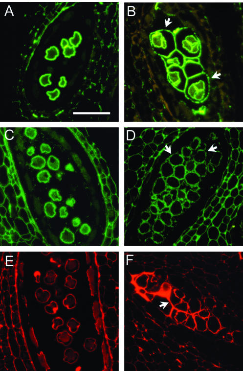
Figure 2.
Immunohistochemical staining of pectin associated with microspores of wild type (A, C, and E) and the qrt3-1 mutant (B, D, and F). Sections were stained with antibodies against unesterified pectin (A and B), methyl-esterified pectin (C and D), and RGII (E and F). In the wild type, the developing pollen grains are separated from each other. In the qrt3 mutant, the developing pollen are enclosed by residual pollen mother cell wall (arrows). Scale bar = 50 μm. The wild-type images were previously published by Rhee and Somerville (1998).

