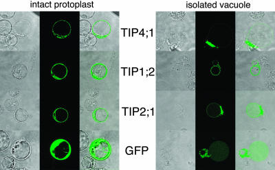
Figure 3.
Subcellular localization of GFP-fused TIPs. Transmission image (left), fluorescence image (middle), and fluorescence superimposed over the transmission image (right). A, GFP-derived fluorescence from protoplasts that were transformed with GFP alone or GFP fused C-terminally to AtTIP4;1, AtTIP2;1, or AtTIP1;2. B, Green fluorescence was also assayed after disruption of the protoplast plasma membrane by osmotic shock using 100 mm Na2HPO4 and 10 mm EDTA, pH 6.5.

