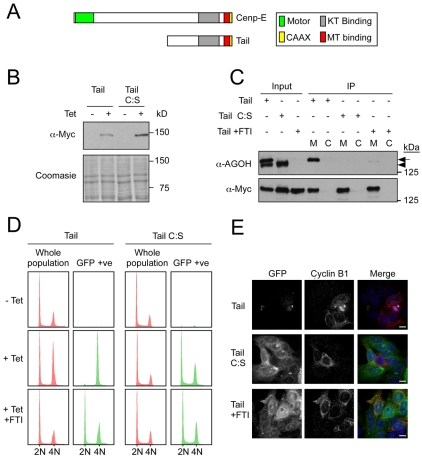
Fig. 4.
Turnover of the Cenp-E tail is farnesylation-dependent. (A) Schematic of Cenp-E showing the tail domain. (B) Immunoblot showing the tetracycline-induced expression of GFP-tagged Cenp-E tail and tail C:S in DLD1 cells. Coomassie blue staining is shown as a loading control. (C) Anti-Myc immunoprecipitates of GFP—Cenp-E tail, GFP—Cenp-E tail treated with FTI and GFP—Cenp-E tail C:S from cells treated with AGOH. The precipitates were then immunoblotted with anti-Myc and anti-AGOH, as indicated. Arrow indicates Myc-tail protein. Arrowhead indicates an unknown farnesylated endogenous protein. M, Myc IP; C, control IP. (D) DNA content of asynchronous DLD1 cultures showing either whole cell populations (red) or only the GFP-positive cells. (E) Immunofluorescence of asynchronous cells expressing GFP—Cenp-E tail, GFP—Cenp-E tail C:S and GFP—Cenp-E tail treated with FTI, then imaged to detect GFP and cyclin B1 (red). Scale bars: 10 μm.

