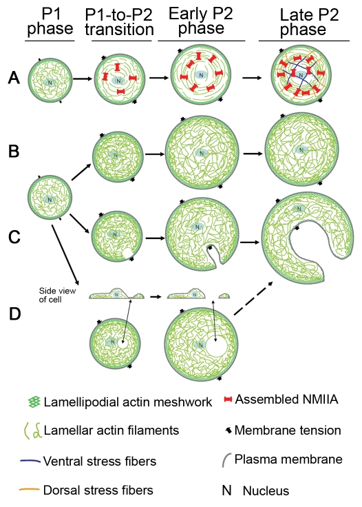
Fig. 7.
Model for development of cytoskeletal coherence and cytoplasmic fragmentation. The P0 phase of early cell spreading is a basal phase and not covered here. (A) There is little NMIIA assembly and contractility and consequent formation of contractile circumferential actin bundles in the fast-spreading P1 phase. As cells approach slow-spreading P2 phase, the assembly and activity of NMIIA increase dramatically. NMIIA contracts and crosslinks actin filaments into a network with tension that prevents cell cytoplasm from being broken by inward plasma membrane tension force. Cells also gain the ability to transmit traction forces from one side of the cell to the other side of the cell. As cell spreading continues through P2 phase, this coherent actin-NMIIA network is expanded. Stress fibers are formed at late P2 stages. (B) The coherent actomyosin-II network is not generated without NMIIA activity. Cells are left with a loose actin network that bears all the inward force exerted by membrane tension. Cell-edge regions with normal levels of actin polymerization are able to resist the pressure of the plasma membrane and do not collapse inward. (C) When peripheral actin polymerization is decreased even from normal variations in activity, the regions with the lowest level of actin polymerization are the weakest points and are most likely to collapse under membrane tension force. These are the initial membrane fragmentation site(s) during the formation of ‘C’- or dendritic-shaped cells. (D) If the regions in cell center are depleted of actin filaments due to lack of actin polymerization and/or a high level of actin depolymerization, holes in the center of the actin network develop. The dorsal and ventral plasma membranes then might come close and fuse, leading to the formation of complete holes. These holes might expand under membrane tension force and eventually also result in the formation of ‘C’ or dendritic shapes.

