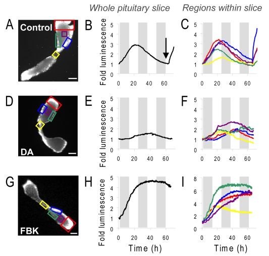
Fig. 2.
Real-time luminescence imaging of PRL promoter-directed transcription in living transgenic pituitary tissue slices. (A,D,G) Luminescent images of 400 μm thick pituitary tissue slices, (B,E,H) graphs of luminescent photon flux from whole pituitary slices and (C,F,I) selected regions within the slices over approximately 3 days, with no treatment (control; A-C), 1 μM dopamine (D-F), or 5 μM forskolin and 0.5 μM BayK8644 (FBK; G,H,I). Arrow in B shows time of stimulation with 5 μM forskolin. Grey bars indicate 12-hour periods. Scale bars: 500 μm.

