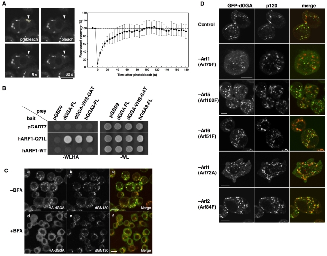
Fig. 3.
ARF-dependent Golgi localization of dGGA. (A) The kinetics of association of EGFP-dGGA with the Golgi were studied in stably transfected S2 cells by fluorescence recovery after photobleaching (FRAP, left panels). Quantification of the FRAP data from a set of three experiments is shown in the right panel. Values are the mean ± s.d. (B) Interaction between dGGA and human ARF1 was examined by a yeast two-hybrid experiment. (C) S2 cells stably expressing HA-dGGA were treated with 2 μg/ml BFA for 1 minute and co-stained with anti-HA (Ba,d, green in Bc,f) and anti-dGM130 (Bb,e, red in Bc,f) antibodies. Scale bars: 10 μm. (D) Effect of ARF protein depletion on EGFP-dGGA localization. S2 cells stably expressing EGFP-dGGA (left column) were mock-treated or treated with dsRNA for ARF79F, ARF102F, ARF51F, ARF72A and ARF84F. They were stained with anti-p120 antibody (middle column). Merged images are shown in the right column (green, EGFP-dGGA; red, p120). Scale bars: 5 μm.

