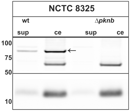
Figure 1. Release of PknB into the growth medium of S. aureus.
The S. aureus strain NCTC 8325 (wt) or a ΔpknB derivative were propagated at 37°C in TSB and harvested at OD600 2. Crude extracts (ce) and supernatant (sup) fractions were isolated, corrected for OD and separated by NuPAGE electrophoresis (Invitrogen). Two-fold higher amounts of the supernatant fractions were used for PAGE as compared to the crude extracts. Immunoblotting was conducted using specific antibodies against PknB (upper panel) or TrxA (lower panel). The latter served as an indicator for cell lysis. The position of the specific PknB signal is marked with a black arrow. The band at ∼60 kDA corresponds to an unidentified protein, which cross-reacts with the antibodies against PknB. The molecular weight of marker proteins is indicated on the left.

