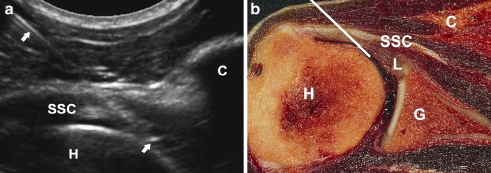
Fig. 5.
US-guided anterior approach. a Sonogram of a right-hand shoulder showing the needle track (arrows) from lateral to medial with the USa approach. The needle is inserted at the level of the coracoid (C). The tip of the needle is in intra-articular position with the tip underneath the subscapular tendon (SSC) and bordering the humeral head (H). b Corresponding cadaver section showing the optimal needle track (white line)

