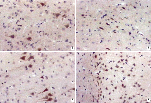
Fig. 5.
Immunohistochemical stain for VEGF of rat brain at 1 and 8 weeks after irradiation, ×400. In acute stage, there are no prominent changes in the radiation group compared to the sham operation group. According to the time interval, the cellularity becomes more prominent and stained cell counts are increased markedly in the white matter of the radiation group. (A) 1 week in the control group, (B) 1 week in the radiation group, (C) 8 weeks in the control group, (D) 8 weeks in the radiation group.

