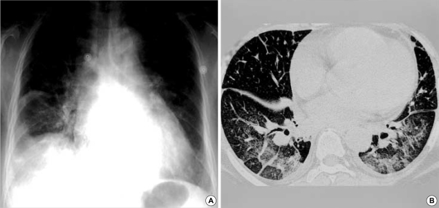
Fig. 2.
Chest radiograph and thin-section CT in a 58-yr-old woman with scrub typhus. (A) Chest radiograph shows diffuse bronchial wall thickening, diffuse ground glass opacities, mild cardiomegaly, bilateral pleural effusions and subsegmental atelectasis. (B) CT of lower zones shows interlobular septal thickening, bronchial wall thickening, diffuse ground glass opacities and patchy consolidations in the dependent lung zones, increased vascular diameter, mild cardiomegaly, bilateral pleural effusions and subsegmental atelectasis.

