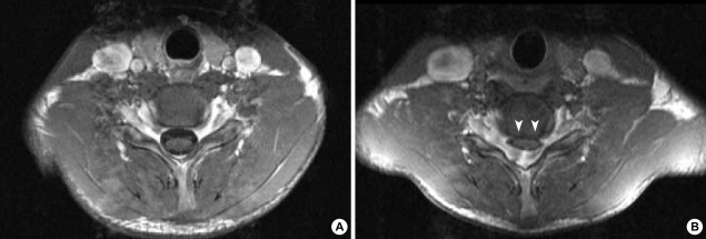
Fig. 3.
(A) The T1-weighted axial image of the sixth cervical cord segment in the neutral position has a normal appearance. (B) There is a striking forward compression by the posterior dural sac in the enhanced axial image of the same cervical cord segment in the anteflexed position (white arrowheads).

