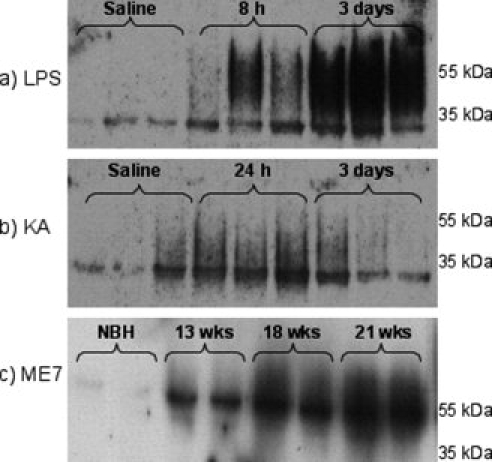
Fig. 2.

Western blot analysis of uPAR expression in hippocampal homogenates. Membrane fraction samples (25 μg protein) were separated by SDS–PAGE, transferred to PVDF, immunoblotted with a polyclonal antisoluble mouse uPAR antibody at 1 μg/mL and developed with ECL. (a) LPS (8 or 72 h) or saline treatment (8 h) (n = 3 for all groups). (b) Kainate (24 or 72 h) and saline groups (24 h) (n = 3 for all groups). (c) ME7 (13, 18, and 21 weeks) and NBH (21 weeks) (n = 4 for each group: one representative gel shown.).
