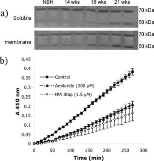
Fig. 3.

Zymographic and 96-well soluble assay of plasmin activity in ME7 animals. (a) Plasmin activity was assessed in both soluble and membrane fractions by in-gel zymography. The activity of both tPA and uPA are visible on the gel at MWs of ∼67 and 50 kDa, respectively (pro forms). Samples are in duplicate and one representative gel of two performed (n = 4) is shown. Images have been captured by digital camera and reversed to increase the clarity of the banding pattern. (b) Plasmin activity was assessed by 96 well assay using ChromazymPL as substrate. Data represent the mean ± SEM for n = 4 in each experimental group. Selective inhibition of plasmin activity at 18 weeks postinoculation with ME7 prion disease using selective inhibitors of tPA (tPA stop, 1.5 μm) and uPA (amiloride, 0.2 mM) revealed the presence of both activities in this membrane fraction.
