Fig. 5.

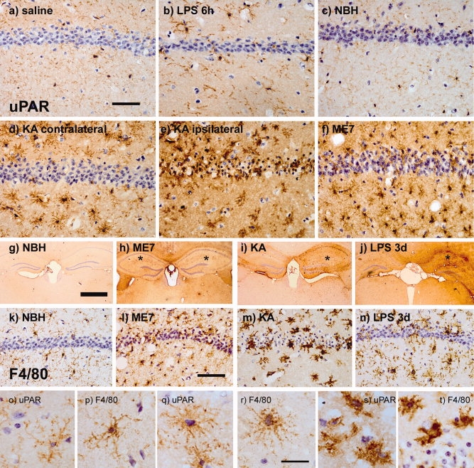
Cellular localization of uPAR expression. PLP-fixed brain sections were immunostained for uPAR (a–j) to identify the cellular source of uPAR in the brain after various insults and for F4/80 (k–n) to confirm morphology and location of macrophages/microglia following these insults. Low-level staining of highly ramified microglial processes can be observed in control animals; (a) saline (8 h) and (c) NBH (18 weeks). Increased expression is evident in (b) LPS (8 h) tissue and (d) contralateral to KA challenge, though the morphology remains quite ramified. Marked expression of uPAR is evident in amoeboid microglia ipsilateral to KA challenge (e) and in condensed microglia in ME7-treated animals (f). In addition to cellular staining, uPAR appears to be released into the parenchyma after ME7 (h), KA (i), and LPS (j) challenges. The site of injection is indicated by *. F4/80 staining reveals constitutive labelling of ramified microglial cells in NBH controls (k). The levels of F4/80 and cell morphology are altered in ME7 (l), KA (m), and LPS 3 days (n). uPAR and F4/80 are also shown at 100× magnification to illustrate the similar morphologies of the cells positively labeled for these antigens (o–t). Both markers display the ramified (saline: o, p), hyper-ramified (kainate contralateral: q, r) and activated/amoeboid (kainate ipsilateral: s, t). Scale bars = 70 μm (a–f), 1 mm (g–j), 50 μm (k–n), and 10 μm (o–t).
