Figure 5.
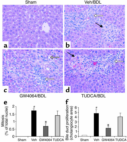
Protection against bile duct ligation–induced necrosis, mitosis, and bile duct proliferation by GW4064. Rats (n = 6) were subjected to bile duct ligation or laparotomy without bile duct ligation (sham-operated). Beginning 24 hours after bile duct ligation surgery, the rats were treated for 4 days with vehicle, GW4064, or TUDCA. Livers were taken for histological analysis 4 hours after the final dose. The panels show representative H&E-stained liver sections from each treatment group at ×400 magnification. (a) Sham-operated rats showing normal liver histology. (b) Vehicle-treated BDL rat showing bile duct proliferation (open arrow) and parenchymal necrosis (filled arrow) with inflammatory cell infiltration. (c) GW4064-treated BDL rat showing bile duct proliferation (white arrow) and fatty cell degeneration (shaded arrow). (d) TUDCA-treated BDL rat showing parenchymal necrosis (filled arrow) with inflammatory cell infiltration. (e) Mitotic nuclei were counted in samples from all rats. Mitosis was quantified by expressing the number of hepatocytes showing mitotic nuclei as a percentage of the total number of hepatocytes. White bars, sham; black bars, vehicle (Veh); dark gray bars, GW4064; light gray bars, TUDCA. Values are presented as average ± SEM. Statistically significant differences between the sham-operated and vehicle/BDL groups are indicated (#P < 0.05). Statistically significant differences between the vehicle/BDL group and either the GW4064-treated or the TUDCA-treated groups are also indicated (*P < 0.05). (f) Bile duct proliferation was quantified by measuring the area occupied by cholangiocytes in 40–50 randomly selected fields under ×400 magnification. Groups as in e; statistics as in e.

