Figure 4.
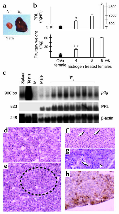
In vivo estrogen induction of PTTG and rat lactotroph tumors. (a) Representative normal rat pituitary (NI) and rat pituitary tumor (E2). (b and c) Serum PRL and pituitary wet weight (b) and Northern blot analysis (c) of pituitary tissue extracts derived from estrogen-treated rats. β-Actin was utilized as the internal control. Ovx, ovariectomized controls. M, marker lane. *P < 0.001; **P < 0.01. (d and e) Reticulin fiber staining (broken circle) of rat anterior pituitary tissue at 24 hours (d) and 1 week (e) after commencement of estrogen infusion. (f and g) Reticulin stain (arrows) (f) and hematoxylin and eosin stain (g) of rat anterior pituitary tissue 4 weeks after estrogen infusion began. Widespread vacuolation, vascular lakes (g, arrow), nuclear pleomorphism and frequent mitosis (g, arrowhead) are visible. (h) pituitary bFGF immunoreactivity after 4 weeks of estrogen treatment. Original magnification, ×200. Reproduced with permission from Nature Medicine (37).

