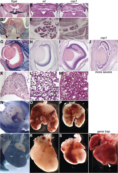
Figure 8.
Additional phenotypic abnormalities in csp1 and Prdm16Gt683Lex mutants correlate with Prdm16 expression. Prdm16 is expressed in choroid plexi epithelium (A), and csp1 mutants exhibit dramatic choroid plexi hypoplasia (B and C). Prdm16 is expressed in the developing salivary glands (black arrowheads in D), and csp1 mutants have severe salivary gland hypoplasia (E and F). csp1 mutants exhibit abnormal retinal folds of variable severity (H–J). Although in situ hybridization shows that endogenous Prdm16 is expressed in the retina in increasingly high levels as embryos progress to birth (data not shown), the Prdm16 gene trap reporter expression is only very weak in the retina, and strong in the pigmented layer of the retina (rpe) (G). We cannot explain this inconsistency. We observe histologic abnormalities in lungs of late embryonic (E19.5) mutants embryos (L and M) and general lung hypoplasia (O and P) when compared with wild-type embryos accompanied by Prdm16 expression in the lung and airway epithelium (K and N). Prdm16 expression in the heart is primarily restricted to the left ventricle as early as E10.5 (N, E13.5 ventral view) and gradually becomes expressed throughout the ventricles, evident postnatally (Q, P3 shown). This expression pattern correlates with gross cardiac ventricular hypoplasia (S), which is more severe in Prdm16Gt683Lex mutants that can exhibit a severe cleft between the ventricles as shown (white arrowhead in T). Lu (lung), LV (left ventricle), RV (right ventricle).

