Figure 4.
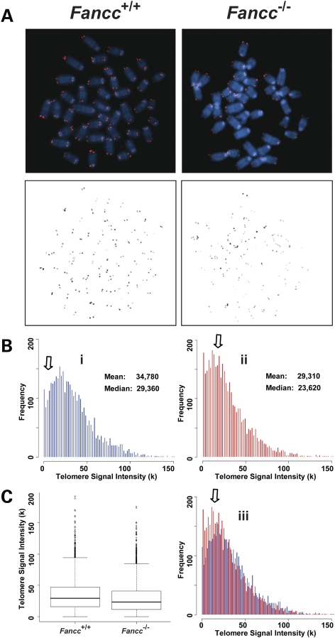
Accelerated telomere attrition is observed in Fancc−/− bone marrow cells after two serial bone marrow transplantations. Q-FISH analysis of wild-type and Fancc−/− bone marrow cells after two serial bone marrow transplantations (n = 4). (A) Representative metaphase spreads of wild-type and Fancc−/− bone marrow cells showing DAPI staining (upper panel, blue) and telomere fluorescence signals (upper panel, red; lower panel, black). There was a decrease in telomere signal intensities in Fancc−/− cells (Bii) in comparison to wild-type cells (Bi), shown here as shift of the dynamic range in overlapping histogram (Biii) and box-plot (C).

