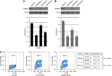
Figure 2.
Effect of MRGBP shRNA on the proliferation of colorectal cancer cells. (A) SW480 and (B) HCT116 cells were treated with MRGBP shRNAs or EGFP shRNA (control) for 48 h, and western blot analysis was performed. Expression of β-actin served as a control. Viability of cells transfected with shRNAs was measured by cell proliferation assay kit. The data represents mean±s.d. from five independent transfections. A significant difference was determined by Student's t-test; *, P<0.05; ***, P<0.001, vs EGFP shRNA-transfected cells. (C) SW480 and HCT116 cells were treated with control or MRGBP siRNA for 48 h, and then were incubated with 10 μM EdU for 30 min. Representative flow cytometric results of HCT116 cells without EdU incorporation (left) and the cells transfected with control siRNA (middle) or MRGBP siRNA (right) were shown. The data represents mean±s.d. from three independent transfections. A significant difference was determined by Student's t-test.

