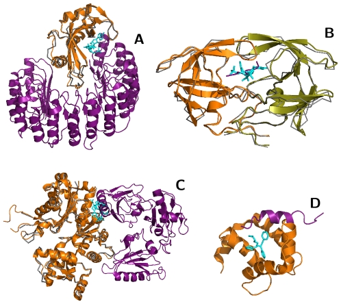
Figure 3. Small molecule binding sites overlapped with four broad classes of protein–protein interfaces.
(A) Enzyme – protein inhibitors: eg, 3′-phosphothymidine (3′–5′)-pyrophosphate adenosine 3′-phosphate (PDB 1U1B:PAX) overlapped with the ribonuclease (orange, 2Q4G)–inhibitor (purple, 2Q4G) interface. (B) Enzyme–protein substrate: eg, Kni-577 (cyan, 1MRW:K47) bound to the HIV-protease dimer (grey backbone, 1MRW:A,B; orange, 1A8K:A,B) at the same positions as its peptide substrate (purple, 1A8K:C). (C) Structural or regulatory interfaces: eg, kabiramide-C (cyan, 1QZ5:KAB) bound to Actin (grey backbone, 1QZ5:A; orange, 1H1V:A) at the same position as Gelsolin (purple, 1H1V:G). (D) Several ligands complemented protein interfaces: eg, bepridil (cyan, 1lxf:BEP) bound at the interface between troponin C (orange, 1LXF:C) and troponin I (purple, 1LXF:I). Figure produced by PyMOL (http://pymol.org).

