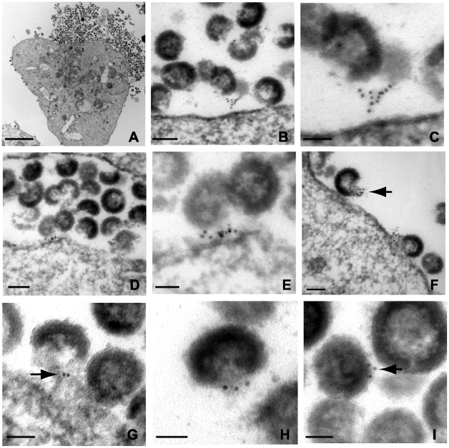
Figure 4. Immunoelectron micrographic analysis of tetherin of NL4.3/Udel-infected A3.01 cells treated with indinavir; labeling at site of particle budding.
(A) Low magnification image of infected cells exhibiting accumulation of immature tethered virions, bar = 2µm. (B) Immunogold labeling of tetherin localized to particle budding sites on the surface of IFN-α stimulated cells. Bar = 100nm. (C) Higher power view from plasma membrane region exhibited in (B); bar = 50nm. Tetherin-positive filamentous structures are shown connecting immature particle to the plasma membrane. (D) Immunogold labeling of plasma membrane region exhibiting accumulation of tethered immature virions and significant labeling of membrane budding site. Bar = 100nm. (E) Higher power view of membrane proximal region from (D); bar = 50nm. HIV-1 virions attached to the plasma membrane exhibit significant tetherin immunolabeling. (F) Tetherin immunolabeling at virion budding locations along plasma membrane. Arrow indicates cluster of tetherin immunolabeling on particle membrane, at “open” end of virion shell. Bar = 100nm. (G) Higher magnification view of tetherin-positive staining at particle budding site. Bar = 50nm. (H) Additional view of tetherin immunolabeling on “open end” of immature virion. (I) Immunolabeling of tetherin associated with virion lipid envelope, bar = 50nm.

