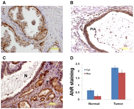
Figure 6. AhR is upregulated in malignant prostate cells.
Prostate sections were immunostained with AhR antibody. Photomicrographs show strong AhR expression in basal cells (arrows in A) and proliferative inflammatory atrophy (B). C. Higher AhR expression is noted in malignant glands (T) compared with normal glands (N). D. Normal and malignant glands were scored using a 0–3 scale. Mean values ±S.E.M. are shown (p-value<0.001, Mann-Whitney test).

