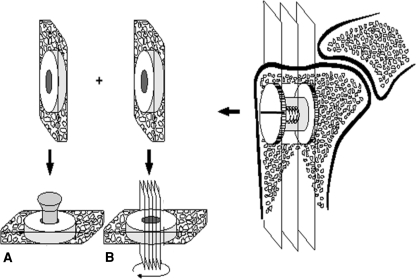
Fig. 2A–B.
A schematic diagram shows specimen preparation. Each bone-implant specimen is cut into two pieces: (A) a 3.5-mm specimen for the mechanical pushout test and (B) a 6.5-mm specimen for histomorphometric analyses. The 6.5-mm specimen is rotated randomly around the long axis of the implant after which four parallel sections are cut parallel to the long axis of the implant.

