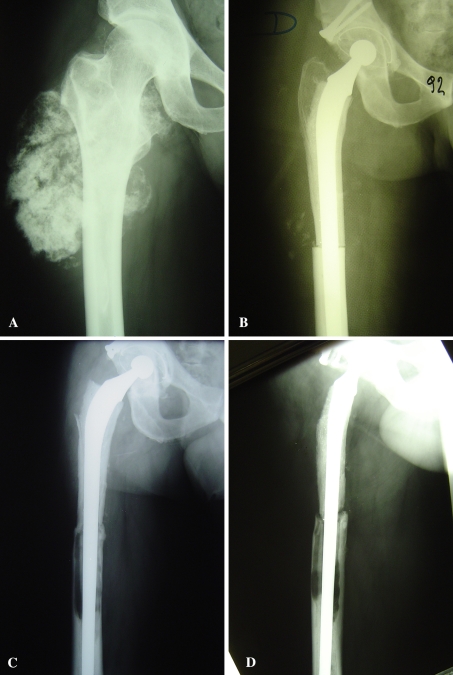
Fig. 2A–D.
(A) This radiograph shows an allograft-composite prosthesis after resection of a chondrosarcoma of the right proximal femur. (B) The immediate postoperative and (C) 16-year postoperative AP and (D) lateral radiographs show allograft resorption, allograft-host bone nonunion, and stem loosening with time. Some mismatch between the allograft and host femur can be seen on the postoperative radiograph.

