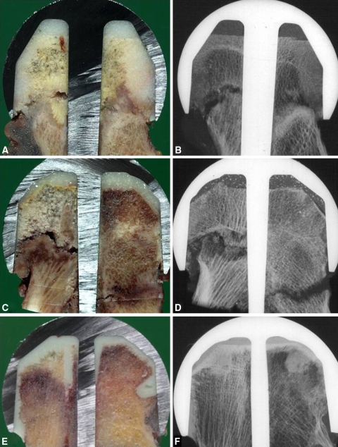
Fig. 3A–F.
(A) A large area of ON after hip resurfacing arthroplasty shows propagation of the fracture line throughout the bordering fibrosis and ON lesion. (B) A corresponding contact radiograph reveals typical bordering fibrosis. (C) An asymmetric segmental ON lesion was observed in hips with partial collapse and a contralateral irregular zone of fibrocartilaginous tissue. (D) A contact radiograph shows several bone fragments in the collapsed tissue and contralateral nonunion. (E) A small triangle-shaped ON lesion with its base at the dome and one side touching a thin deep additional fixation drill hole is seen proximally. (F) Sclerosis is evident at the border of the triangle-shaped ON lesion.

