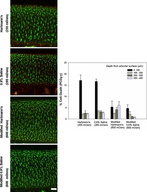
Fig. 4.
Coronal CLSM projections of the injured cartilage edge and PCDFT compare the extent of cell death between 0.9% saline and Hartmann’s solution. The panels show coronal CLSM projections of the full thickness of articular cartilage. The bar chart shows the corresponding pooled data for PCDFT as a function of increasing depth from the articular surface. In situ chondrocyte death is localized mainly near the articular surface (ie, superficial zone, 0- to 100-μm depth interval) for explants exposed to control solutions of Hartmann’s solution (255 mOsm) and 0.9% saline (285 mOsm) with relative sparing of the middle and deep zones. For explants exposed to the modified, high osmolarity (600 mOsm) of Hartmann’s solution and 0.9% saline, there is a decrease in PCDFT in the 0- to 100-μm depth interval of injured articular cartilage (N = 3, n = 12, white bar = 100 μm). CLSM = confocal laser scanning microscopy; PCD = percentage cell death.

