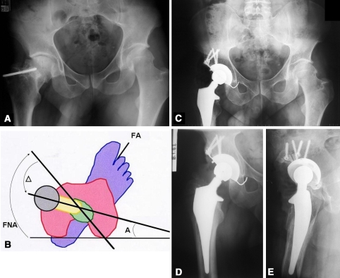
Fig. 2A–E.
(A) A preoperative frontal view of the pelvis shows left hip osteoarthritis. The patient was a 49-year-old woman who subsequently underwent a left THA. (B) The values for femoral neck anteversion (FNA = 50°), the desired final prosthetic anteversion (anteversion, A = 15°), and the correction made in the prosthetic neck (∆ = −35°) are shown. FA = foot axis (25°). A postoperative (C) AP view of the pelvis. (D) AP and (E) mediolateral views of the hip obtained at the 9-year followup show satisfactory leg length equalization and femoral offset restoration.

