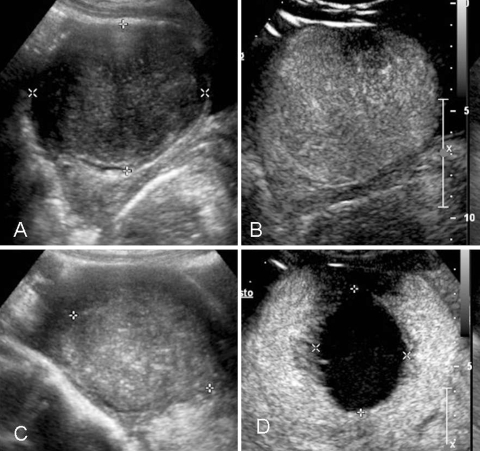
Fig. 3.
A 41-year-old woman affected by a 7-cm symptomatic uterine fibroid. A B-mode US shows a hypoechoic mass in the uterus. B CEUS confirms rich vascularization inside the mass. C B-mode US 6 months after percutaneous RFA: the treated fibroid appears nonhomogeneous and reduced in size. D CEUS confirmed a huge area of necrosis with a peripheral rim of enhancement in a normal uterus without any residual disease

