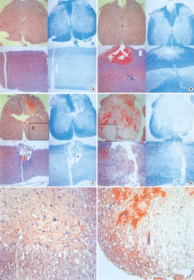
Fig. 6.
Representative coronal and sagittal sections of the lesions 14 days after the injury. The dorsal surface is at the top. The left panels are hematoxylin and eosin stain and the right are Luxol fast blue stain (Original magnification, ×40). The Luxol fast blue stain clearly reveals residual myelinated white matter that is restricted to a peripheral rim in the more severe injury groups. (A) control, (B) Group 1, (C) Group 2, (D) Group 3, (E, F) Higher power magnification of the ventral-lateral funiculus (the block demarcates the region of spared white matter, ×100). *: Cystic formation. (E, F) Higher power magnification of the ventral-lateral funiculus (the block demarcates the region of spared white matter, ×100).

