Fig. 1.

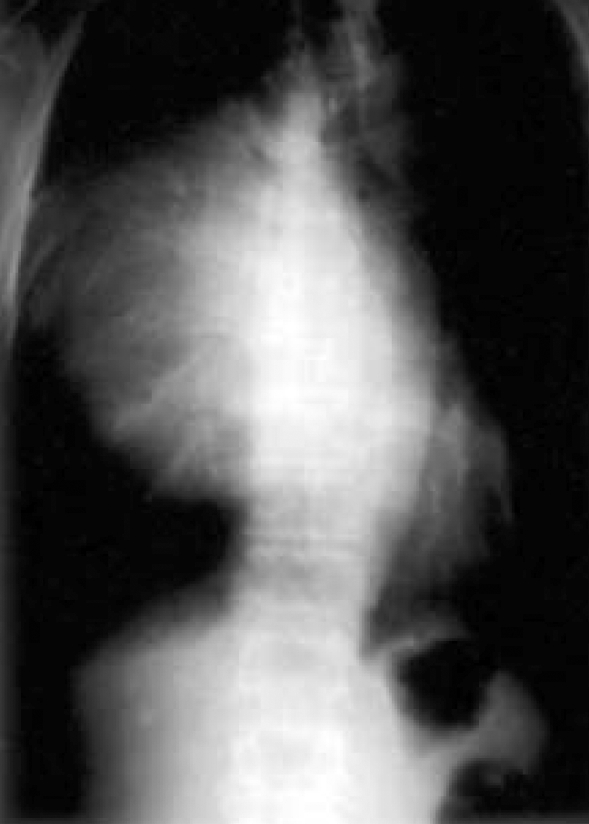
PA radiographs of the chest shows a huge, well marginated, homogeneous mass in the anterior mediastinum displacing the lower trachea, bronchus and heart contralaterally.

PA radiographs of the chest shows a huge, well marginated, homogeneous mass in the anterior mediastinum displacing the lower trachea, bronchus and heart contralaterally.