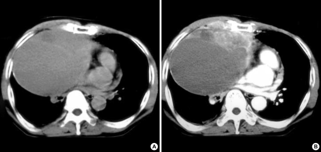
Fig. 2.
(A) Chest CT scan at the level of aortic origin demonstrates a huge, low attenuating, cystic mass in anterior mediastinum with internal septa and heterogeneous solid area anteromedially. (B) Contrast enhanced CT scan shows the focal solid mass area with heterogeneous enhancement anteromedially and irregular margin between the mass and anterior chest wall and pericardium.

