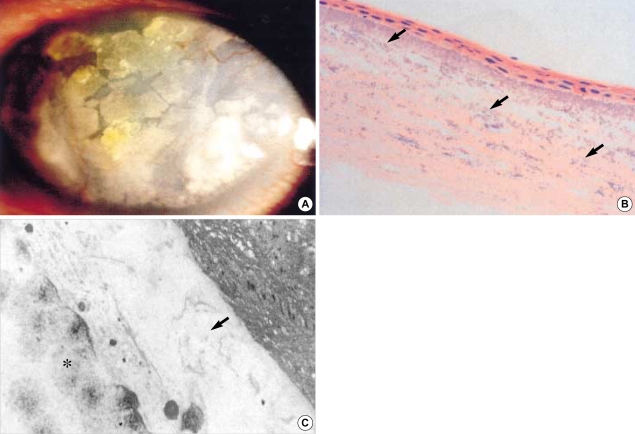
Fig. 1.
(A) Calcium is deposited on the cornea (visual acuity: light perception). (B) Multiple calcium impactions and calcium deposit are observed in the stromal layer (H&E, ×200). (C) Deposited calcium is seen in stromal layer (asterisk). Destroyed basement membrane is observed in the degenerated stromal space (arrow) (×4,100).

