Figure 6.
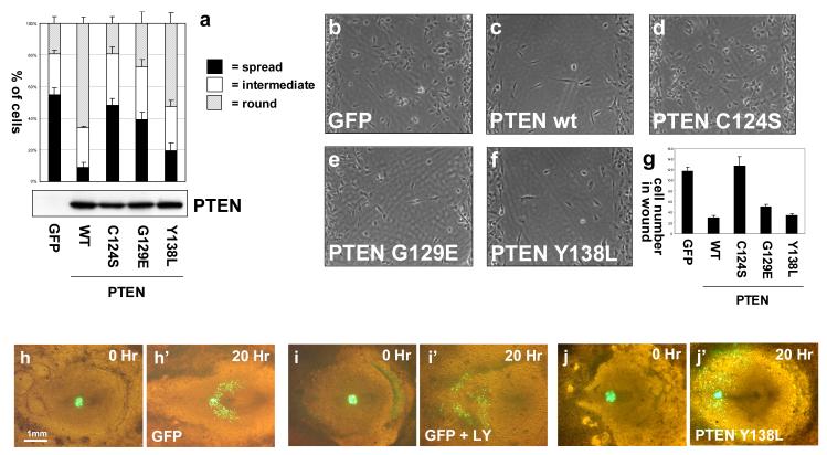
The regulation of cell spreading, and cell migration by PTEN. (a) Trypsinised U87MG cells expressing GFP or different PTEN proteins were allowed to adhere onto collagen IV coated culture slides for 30 minutes before fixation. Data are presented as the mean fractions of each cell population blind scored as spread, intermediate or round, + SEM from 5 randomly selected fields from a representative experiment. This experiment has been repeated twice on collagen and twice on plastic with similar results. Equal expression of the PTEN mutants was verified by Western blotting and the same set of transduced cells was used in the following experiments addressing adgherent cell migration. (b-f) Trypsinised U87MG cells expressing GFP or different PTEN proteins were seeded at confluence and allowed to adhere to matrigel-treated plastic before the culture inserts were removed to leave a defined 500μm cell-free gap. Cells were then allowed to migrate over a 24 hour period, before cells were fixed and photographed. (g) Quantitation represents the mean number of cells in a central area of the gap from 6 randomly selected fields +/− SEM. This experiment was performed three times with similar results. (h-j) PTEN Y138L interferes with directed cell migration in the developing chick embryo, but does not block cell motility. Cell migration assays were performed using cells expressing GFP or GFP-PTEN Y138L as previously described (Leslie et al., 2007; Yang et al., 2002).

